Routine Quantitative Image Analysis (Q.I.A)
A range of software modules for more than 15 markers in IHC, FISH, double labelling...
- Automated detection of all regions of interest and all cells for accurate quantification.
- Virtual slides classification and management.

Benefits
Objective quantitative results
Completely automated, transparent for the user, the detection of tumor areas, cells and quantitative data, provides the user with virtual slides, including the evaluation of a range of routine or emergent markers.
Time saver
Save by over 5-folds personnel time compared to conventional eye screening.
Pathologist or technician spends less time on difficult cases; thus, more time for urgent tasks
Facilitate the training of junior staff.
Speed up report submission.
Integrated to LIMS
Included in the solution (HL7, JSON, direct Data-base supported and integrated web server).
Traceability / Quality assurance
Compliance with regulated environments, including user-rights management, audit trail and support for write-only media.
Image Gallery
PathoScan T-M IHC HER2
Invasive Breast cancer biopsy HER2/IHC and HES staining Dual view Software score 1+
PathoScan T-M IHC HER2
Invasive Breast cancer biopsy
Software score 1+ Pathologists's score 1+
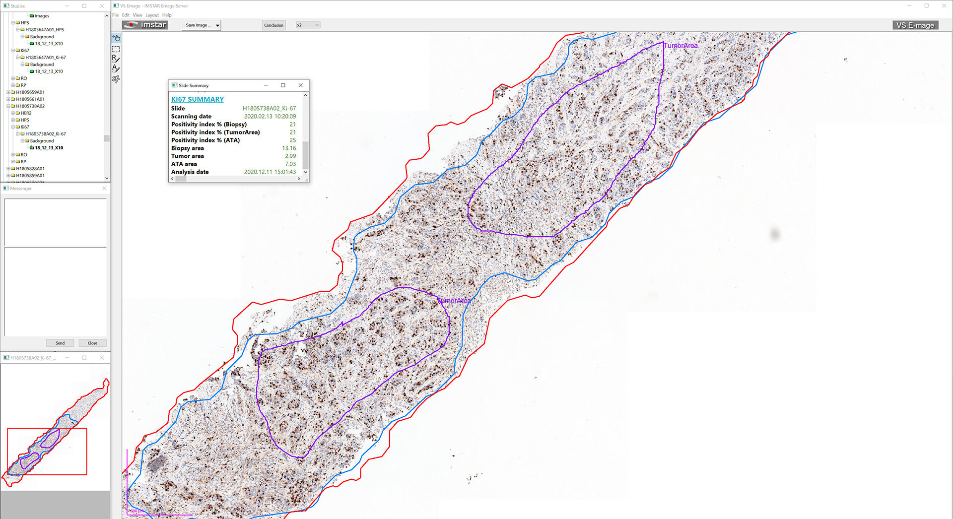
PathoScan T-M IHC Ki67
Invasive Breast cancer biopsy Pathologist's index 25%,
Software index 21%
Eyeballing control (1000 cells) index 22%
PathoScan T-M IHC Ki67
Invasive Breast cancer Ki67/IHC and HES staining Dual view Software Ki67 index 33%
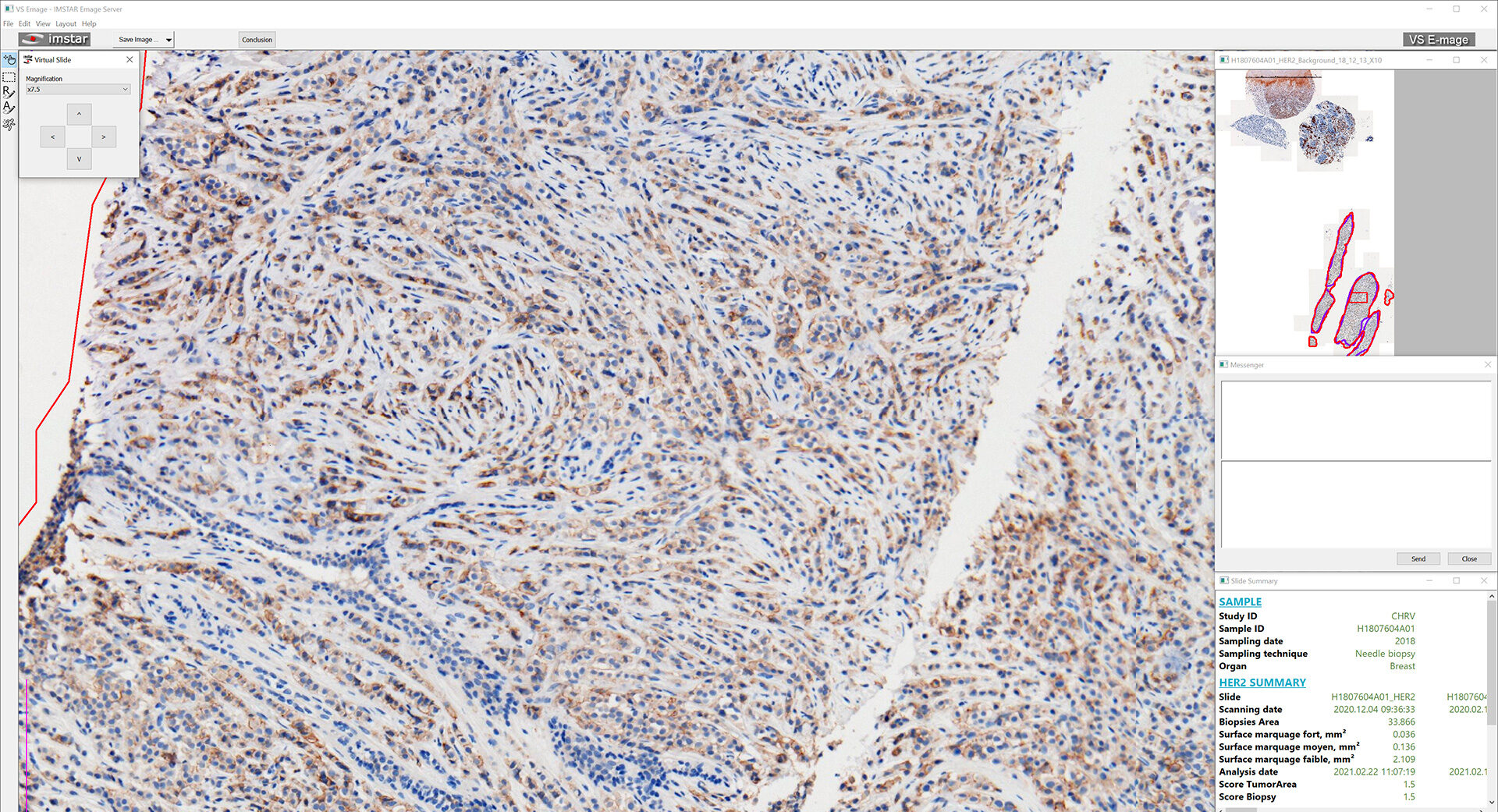
PathoScan T-M IHC HER2
Invasive Breast cancer HER2/IHC and HES staining Dual view HER2 software score 2+
Expert vs QIA processing comparison
Comparison between expert HER2 assessment and the score generated through our proprietary QIA processing. Both scores are concordant (2+)
PathoScan T-M IHC HER2/KI67
Zytomed Double label HER2/Ki67 Software Ki67 index 21% Software HER2 score 3+
PathoScan T-M FISH
Probe: Zytolight ERBB2/CEN17 - Invasive Breast cancer Software ratio 5.7
PathoScan T-M Cervical LBC PAPS
Automated detection of suspicious cells in a gallery for validation by the cytologist, and its position on the virtual slide
More Informations
Posters & Presentations
- [Carrefour Pathologie 2023, Paris, FRANCE]: Immunohistochemical determination of HER2-positive, HER2-low and HER2-ultralow status in breast cancer by quantitative image analysis algorithm. M. Cossutta, JP. Bellocq, C. Egele, A. Papine, E. Tatarinova, K. Socha, M. Soussaline, C. Homsy, F. Soussaline Read more
- [Carrefour Pathologie 2023, Paris, FRANCE]: Automated evaluation of Ki67 index in breast cancer and in neuroendocrine tumor by quantitative image analysis. M. Cossutta, JP. Bellocq, C. Egele, A. Papine, E. Tatarinova, K. Socha, M. Soussaline, C. Homsy, F. Soussaline. Read more
- [35th European Congress of Pathology 2023, Dublin, IRELAND ]: Validation of a Quantitative Image Analysis Algorithm for HER2 Status Determination in Breast Cancer by Immunohistochemistry as Positive, Low or Ultra-Low. M. Cossutta, JP. Bellocq, C. Egele, A. Papine, E. Tatarinova, K. Socha, M. Soussaline, C. Homsy, F. Soussaline. Read more
- [35th European Congress of Pathology 2023, Dublin, IRELAND ]: Validation of a Quantitative Image Analysis Algorithm for
Ki67 Index in Breast Cancer and Neuroendocrine Tumor. M. Cossutta, JP. Bellocq, C. Egele, A. Papine, E. Tatarinova, K. Socha, M. Soussaline, C. Homsy, F. Soussaline. Read more - [19th European Congress on Digital Pathology 2023, Budapest, HUNGARY]: Digital Image Analysis as a Standardized Method for External Quality Assessment of HER2 IHC in Breast Cancer. M. Cossutta, R. Røge, H. L. Kristoffersen, E. Tatarinova, A. Papine, M. Soussaline, S. Nielsen, F. Soussaline
- [USCAP 112th Annual Meeting 2023, New Orleans, USA]: Digital Image Analysis-Aided Assessment of HER2 Immunohistochemistry for External Proficiency Testing. R. Røge, M. Cossutta, H. L. Kristoffersen, E. Tatarinova, A. Papine, F. Soussaline, S. Nielsen. Read more
- [Carrefour Pathologie 2022, Paris, FRANCE]: Automated Evaluation of Ki67 and HER2 in Breast Cancer and Ki67 in NETs by Image Analysis. M. Cossutta, A. Papine, E. Tatarinova, C. Homsy, M. Soussaline, C. Egele, JP. Bellocq, F. Soussaline
- [Carrefour Pathology 2019, Paris, France, November]:Quantification digitale des marqueurs immuno histochimiques du cancer du sein. Évaluation du système d’analyse d’image automatisée IMSTAR Pathfinder Tumor-Marker. Socha K., Glaser C., Bienvenu L., Tatarinova E., Papine A., Soussaline F., Soussaline M. (2019) Read more


